WILLS EYE IS HIRING – CLICK HERE TO APPLY TODAY!

Glaucoma Symptoms and Diagnosis hero
GLAUCOMA SYMPTOMS & DIAGNOSIS

The best way to test for glaucoma is through a comprehensive eye exam with an ophthalmologist. If you have risk factors for glaucoma, you will require not just one exam, but also repeat examinations at an appropriate frequency to evaluate for the development of glaucoma over time.


GETTING A GLAUCOMA DIAGNOSIS

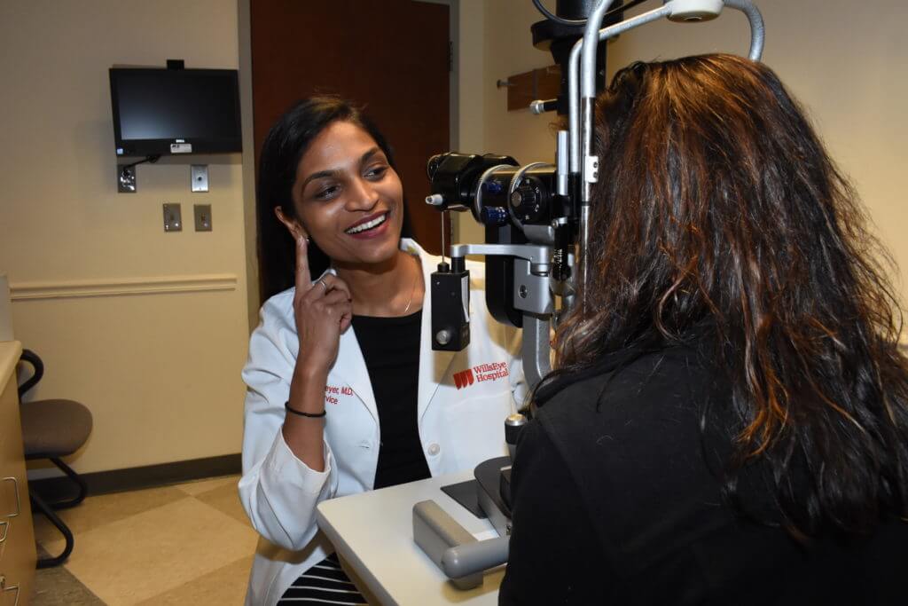
The best way to test for glaucoma is through a comprehensive eye exam with an ophthalmologist. The examination includes an evaluation of your angle (gonioscopy), and your optic nerve with dilation if appropriate. Additionally, a visual field test and optic nerve photograph or analysis may also be performed. If you have risk factors for glaucoma, you will require not just one exam, but also repeat examinations at an appropriate frequency to evaluate for the development of glaucoma over time.

What Are the Signs & Symptoms of Glaucoma?

For most people with glaucoma, there are no warning signs or noticeable changes in vision until the disease has silently and gradually worsened to a severe stage. About half of those with glaucoma do not yet know they have it. This is why glaucoma is often called the “thief of sight” and one of the main reasons why it is still one of the leading causes of blindness in the world.

 
In the early stages, glaucoma typically affects peripheral (or side) vision. A common misconception is that those with glaucoma get tunnel vision. Tunnel vision is almost never the way patients experience their glaucoma. Instead, vision changes start with a very subtle blurring which usually begins near the nose. Since we see with both eyes at the same time, vision lost in one eye is often covered up and hidden by the other eye. If left untreated, areas of vision loss will slowly spread and may go unnoticed until it reaches the central vision.

On the other hand, some people may develop acute glaucoma which often presents with severe vision loss and pain that is brought on rather quickly. Symptoms are usually a combination of:

  • Severe eye pain and headache
  • Blurry vision
  • Redness of the eyeball
  • Nausea (upset stomach) or vomiting

Acute angle closure glaucoma is the most well-known cause of acute glaucoma. Other causes may include blood filling the eye after an injury (hyphema) and certain medications such as topiramate (Topamax) or prolonged steroid use. If you are experiencing these symptoms, please contact your local eye doctor or emergency room as soon as possible.


Tests for Glaucoma - Diagnosis and Monitoring

Generally, glaucoma tests are divided into two groups:

  • Assessing the structure of the optic nerve, and
  • Assessing the function of the optic nerve

The tests below are the typical tests used to diagnosis and monitor glaucoma. There are, however, many other tests that the doctor may request depending on your examination. These tests range from color vision tests to other tests of eye structure, and may include imaging of the eye and brain in some cases.

TESTS OF STRUCTURE OF THE OPTIC NERVE
  • Photography of Optic Nerve. A digital camera takes photos of the back part of the eye for evaluation of the optic nerve. The photos are used to document how your optic nerves look at this exam – these photos can be compared to prior photos or may be used in the future for comparison.
  • Optical Coherence Tomography or OCT (a computerized picture of the optic nerve). This specific image measures the microscopic thickness of the optic nerve fibers and compares the readings with the normal database and report the results in colors and numbers. The numbers will be compared over time with prior and future pictures, to identify if any change has occurred in the structure of your optic nerve.


TEST OF FUNCTION OF THE OPTIC NERVE

Visual Field Test. You will be asked to cover your left eye with a patch and then your chin will be placed in the chin rest area of the visual field machine. The machine looks like a big bowl. Once your head is positioned correctly and the proper lens (based on your glasses prescription) is placed in front of your eye, you will be given a button to press when you see a light flash. You should see a light source in the center of the dome (fixation light). You are to look at the fixation light for the duration of the test.

While looking at the fixation lights, white lights will flash throughout the “dome.”  Every time you see a white light flash, you should push the button. The white lights will start out bright, but become dimmer as the test progresses. They will become very difficult to see – the test is designed this way – it will test not only how far into the periphery you see, but also how sensitive your peripheral vision is to very dim lights.

It is completely normal to miss some flashes so it is best to stay calm even if you feel like you have not seen a light for a long time.