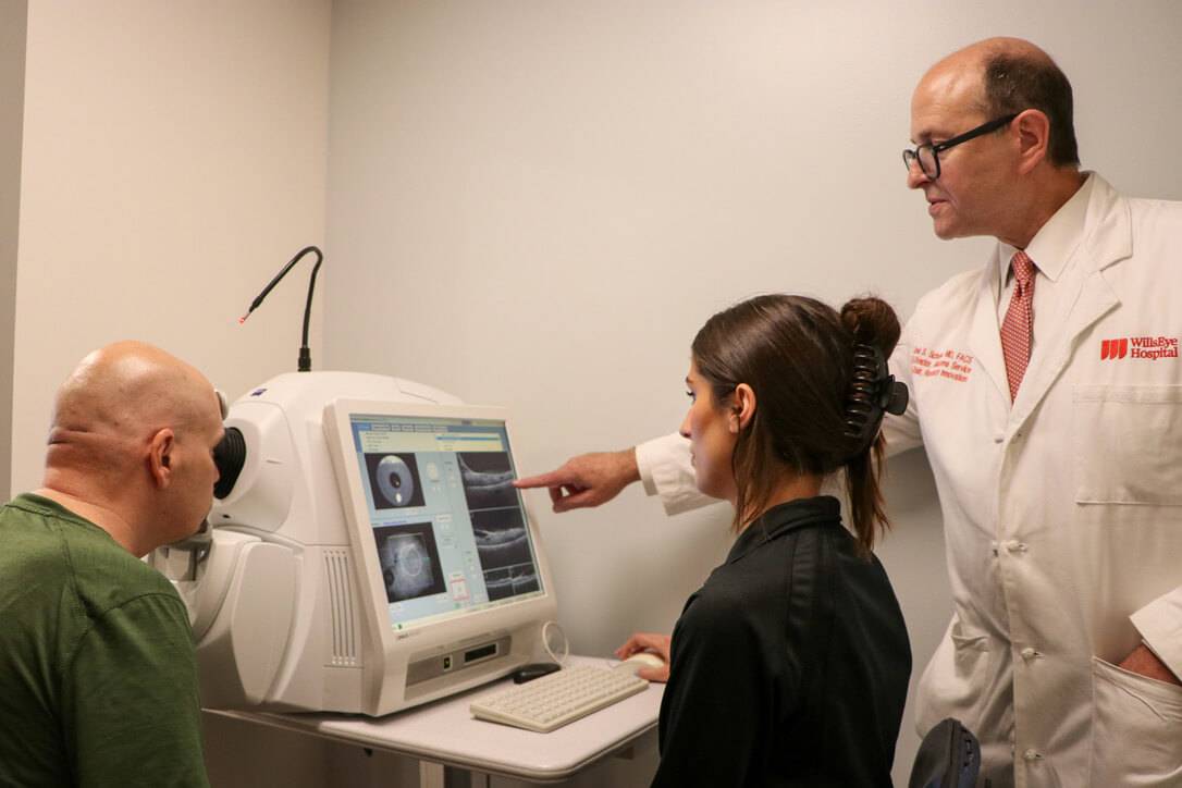
IN NEUROIMAGING (ACORN)


Under the leadership of Co-Directors Joel S. Schuman, MD, FACS and Gadi Wollstein, MD, the Advanced Center for Ophthalmic Research in Neuroimaging (ACORN), is a team of ophthalmologists, engineers, software specialists, statisticians, and trainees that focus primarily on glaucoma research.
Glaucoma, the second leading cause of blindness worldwide, is a neurodegenerative disease that affects the retina and optic nerve of the eye. Although various interventions exist, there is currently no known cure for glaucoma, therefore early disease detection is of the utmost importance for vision conservation.
The diagnosis of glaucoma requires the identification of structural changes in the back of the eye accompanied by evidence of visual field loss. The incorporation of ocular imaging devices as part of the clinical evaluation allows for accurate, micron scale measurements of the structures in the back of the eye. This can improve disease detection and assist in monitoring changes over time.
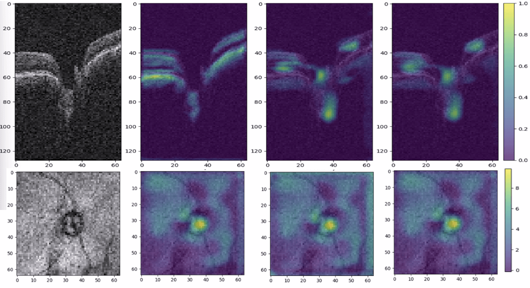
OCT works by using a technique called interferometry. Light is shone on the back of the eye and the machine uses the light that bounces back to create a virtual cross section using the different reflectivity of tissues at different depths. The first OCTs were called time domain (TD)-OCTs, which obtained information about different depths in the tissue by using a moving mirror. Current OCT devices are called spectral domain (SD)-OCTs, which have eliminated the need for a moving mirror by using a mathematical technique called a Fast Fourier Transform and incorporating a spectrometer and charge-coupled-device camera to separate and detect the tissue layer information.
Newer experimental OCT technologies include swept-source OCT, polarization sensitive OCT, phase-sensitive OCT, visible light OCT, and others that further enhance the ability to visualize fine structures in the eye and assess the functionality of certain tissues.

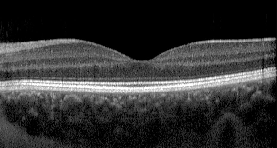
OCT cross-section of the macula

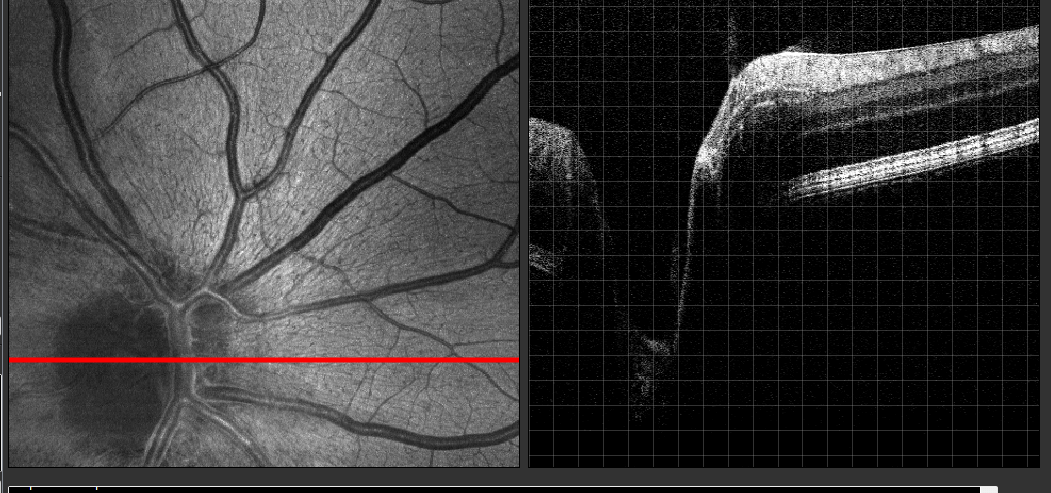
OCT circular scan around the optic nerve region

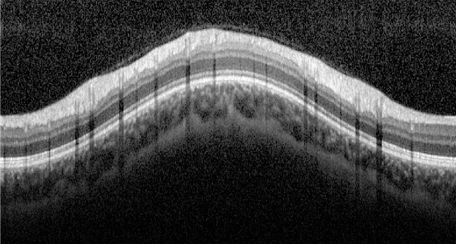
OCT cross-section of the optic nerve head

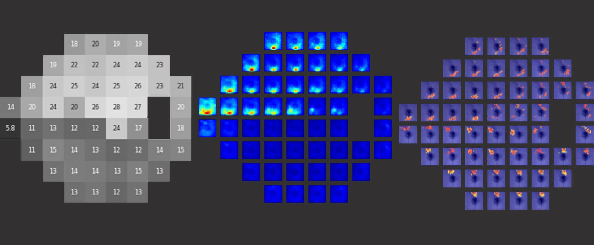
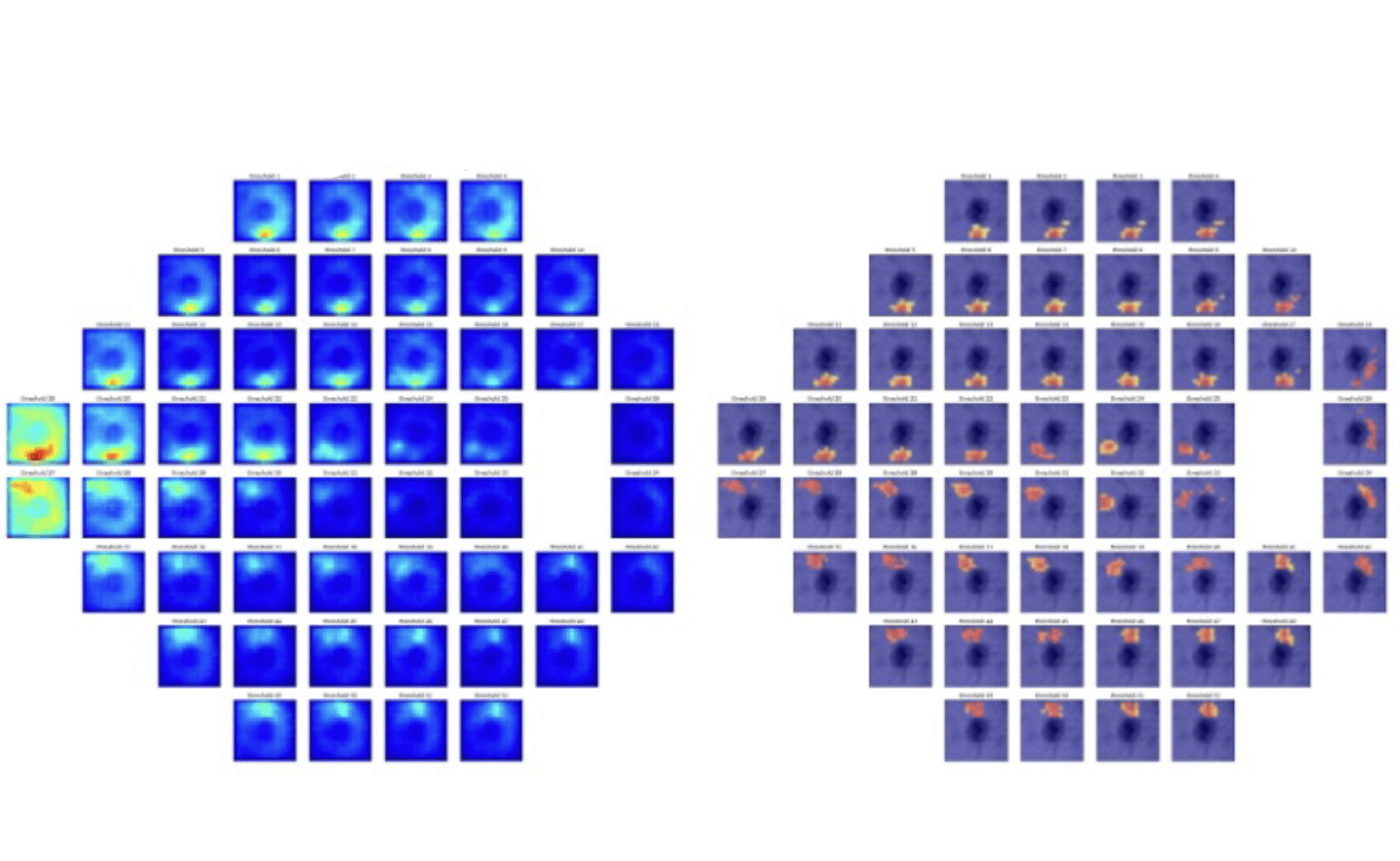
As the neurons that comprise the RNFL die this layer thins, giving ophthalmologists a way to assess the severity of glaucomatous damage. Regions of retinal nerve fiber layer (RNFL) thinning are shown here, marked with red and yellow colors, expanded over the course of six years of follow-up.
Our visionary team is at the forefront of scientific inquiry into pioneering innovations and empowering clinicians and scientists to provide extraordinary care informed by the latest biomedical research.

Prediction of Disease
Early detection of disease is critical to slowing down degeneration of neuroretinal tissue and preserving vision. Our group has pioneered methods for early detection, sensitive monitoring, and precise prediction to improve clinical evaluation and indication for treatment.

Our mission is to delve into the root causes of disease pathogenesis, using advanced imaging and analytical techniques developed in our lab. These discoveries not only improve detection and prediction of disease but also may inform new targets for disease treatment.

Imaging Technology
The ACORN Lab is home to Philadelphia's most comprehensive Ocular diagnostics imaging center. Our lab directors pioneered the development of technologies now considered standard of care in the eye, and continue to push the boundaries of ophthalmic imaging with cutting edge technology.

Methodologies
The age of Artificial Intelligence and computational accessibility have ushered in new and improved methods for understanding disease. Our team is at the forefront of developing statistical and machine learning algorithms for extracting insights from clinical and imaging data to better understand disease features and improve prognostication.


