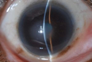
WILLS EYE IS HIRING – CLICK HERE TO APPLY TODAY!

Fuchs' Dystrophy
FUCHS' DYSTROPHY

An abnormality involving guttae on the back layer of the cornea (endothelium). It is generally slowly progressive, leading to mild or severe corneal swelling (edema).

Call Us: 215-928-3180


Fuchs' Dystrophy Slit LampOVERVIEW

Fuchs' dystrophy is a condition where the endothelial cells on the back layer of the cornea are not normal. Healthy endothelial cells are required to keep the cornea clear. Most patients with Fuchs dystrophy have a very mild form that never affects vision. When it does affect vision, it usually occurs in middle age or later. In mild Fuchs dystrophy, the vision may be slightly decreased. As the Fuchs dystrophy progresses and the corneal swelling worsens, the vision slowly declines. It is often worse in the morning than later in the day. Late in the course, pain or severely decreased vision can occur.

Treatment options include:

  • Drops (saline-like drops) to decrease swelling and later corneal transplantation
  • Either a partial thickness corneal transplant (e.g. Descemet’s stripping endothelial keratoplasty [DSEK] or Descemet’s membrane endothelial keratoplasty [DMEK]), or a full thickness corneal transplant (especially in eyes with corneal scarring.) In some patients with fairly mild and central Fuchs dystrophy, removal of the abnormal cells without a corneal transplant (Descemet’s stripping only [DSO]) may be an option.

DEFINITION

An abnormality involving guttae on the back layer of the cornea (endothelium). It is generally slowly progressive, leading to mild or severe corneal swelling (edema).

SYMPTOMS

Initially no symptoms. Then can develop blurred vision, often worse in the mornings, which may clear over a few hours. As the condition progresses, the blurred vision can worsen and last all day long. The swelling can cause painful blisters as the condition progresses.

CAUSES

Often a genetic predisposition and can run in families.

RISK FACTORS

Family history of Fuchs dystrophy

COMPLICATIONS

Corneal scarring can develop in severe or longstanding cases.

TESTS AND DIAGNOSIS

The diagnosis can usually be made during a slit lamp examination. Ancillary testing can include measurement of the corneal thickness (pachymetry) and endothelial cell imaging (specular microscopy).

TREATMENT

Treatment ranges from observation in mild cases to salt drops and ointment (hypertonic sodium chloride 5%) to surgery including partial thickness corneal transplant (e.g. Descemet’s stripping endothelial keratoplasty [DSEK] or Descemet’s membrane endothelial keratoplasty [DMEK]) or a full thickness corneal transplant (especially in eyes with corneal scarring). In some patients with fairly mild  and central Fuchs dystrophy, removal of the abnormal cells without a corneal transplant (Descemet’s stripping only [DSO]) may be an option.

PREVENTION

Routine eye examinations are recommended to monitor the condition. Longstanding swelling increases the chances of developing corneal scarring which decreases the success rate of partial thickness corneal transplantation.